
47: 尿路结石—肾脏手术治疗
阅读本章大约需要 4 分钟。
引言
世界各地形成结石的总体概率不同,估计亚洲为1–5%,美国为10–15%,而全球范围内则根据年龄、性别、种族和地理位置不同在1–15%之间。1 在过去几十年里,全球肾结石的患病率有所增加。在美国,成年人在2007–2010年期间的患病率估计为8.8%。这种结石病的增加在青少年人群中也有报道,其中女孩受结石病影响多于男孩。2,3,4 结石病发病率的上升导致急诊就诊、住院、手术和医疗费用的增加。在美国,儿童尿路结石的医疗费用每年至少达3.75亿美元。5 此外,儿童在首次发生结石后的3至5年内,结石复发率约为50%。6,7,8 然而,多达60%的儿童出现的结石不易自行排出,因此在此类患者中手术治疗仍是主要治疗方式。9 不过,过去几十年间手术治疗经历了范式转变。在过去50年里,儿童肾结石的治疗已从开放手术过渡到ESWL,再到内镜治疗。到20世纪80年代之前,大多数肾结石采用开放手术治疗;然而,自20世纪80年代起,随着ESWL和PCNL首先在成人中的应用,其在儿童人群中的安全性和有效性也得到证实。目前,随着器械的微型化,以PCNL和RIRS为代表的内镜治疗显著提升了对患儿肾盂肾盏系统的进入性,并以最少的并发症实现最高的结石清除率。10
体外冲击波碎石术
体外冲击波碎石术(ESWL)于20世纪80年代初首次用于治疗成人肾结石。在成人中确立其安全性之后,到1986年很快被应用于儿童人群。11,12 最初,由于设备存在技术挑战,制造商仅建议将该治疗用于身高超过135厘米的患者。ESWL 是治疗直径不超过15毫米的肾结石的治疗选择之一。对于直径大于15毫米的结石,与泌尿内镜治疗相比,再治疗率显著升高,结石清除率也降低。
随着碎石机的改进(取消水浴并采用超声引导的结石定位成像),儿童体外冲击波碎石术(ESWL)的接受度和应用逐渐提高。儿童患者的无结石率在65%至71%之间,取决于结石所在部位、能量设置以及所用机型。13
ESWL 的优势
ESWL所提供的最重要优势是这是一种非侵入性治疗。另外,单次或两次治疗即可达到约68%至84%的无石率。这可归因于儿童体积较小,使冲击波衰减较少,且在体内传播更好,从而获得更佳的碎石效果。此外,儿童的输尿管较成人更具顺应性,因此儿童的输尿管比成人群体更容易排出结石。
ESWL 的缺点
与泌尿内镜手术相比,最重要的缺点是结石清除率较低。其次,即便是 ESWL,在绝大多数儿科患者中也需要全身麻醉(图1)。第三,人们担心 ESWL 对处于生长发育中的肾脏的影响。Lifshitz 等 对29例进行了平均9年的随访,发现接受治疗的肾脏生长呈下降趋势,并出现1例高血压。。14 然而,作者认为这些变化是由于肾脏本身异常所致,并非 ESWL 造成。对39名接受 ESWL 治疗的儿童进行6个月至8年的进一步随访,未发现高血压,DMSA 扫描亦未见肾瘢痕。15 Villanyi 等 在16名年龄6至14岁的儿童中观察到,肾功能未见恶化,但各种肾损伤替代指标出现短暂排泄,例如 β2-微球蛋白,持续约一周。因此,建议两次连续 ESWL 治疗之间至少间隔15天。16 然而,目前没有充分证据表明 ESWL 在儿科人群中具有任何显著的长期影响。
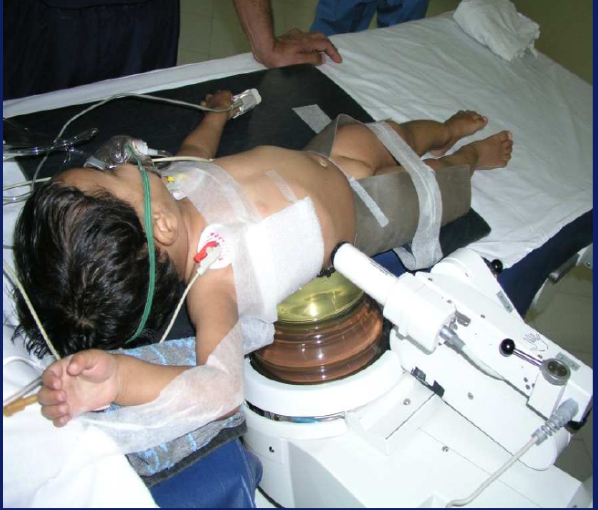
图 1 儿科患者在全身麻醉下行 ESWL
经皮肾镜取石术
PCNL 已经取代了开放手术,目前被认为是直径大于 20 mm 的肾结石的一线治疗方法,结石清除率超过 90%。17
PCNL最初由Johannson和Fernstrom于1976年描述。自那以后,它已成为治疗肾结石的主要治疗手段。1985年有关于儿童PCNL的报道:一项病例系列,纳入7名年龄在5至18岁之间、使用标准成人器械的患者。18 然而,随着光学器件、肾镜尺寸的持续减小,以及更先进的碎石技术的出现,使得PCNL可以使用更小的通道,并降低了并发症发生率。微通道PCNL(通道直径可达20 Fr)、超微通道PCNL(11 Fr、13 Fr)、迷你-微通道PCNL(8 Fr)、微型PCNL(鞘管直径4.85 Fr)等,使得即使在更年幼的儿童中,人们对PCNL的接受度也更高。
PCNL在全身麻醉下进行。首先行膀胱镜检查,并将4 Fr或5 Fr输尿管导管置入肾盂肾盏系统。它用于PCS的显影与扩张以及术中冲洗。随后将患儿置于俯卧位。初始穿刺可在超声引导或透视引导下进行。由于儿童肾脏较成人肾脏更具活动度,初始穿刺是手术中最重要的部分。儿童肾脏在初始穿刺和扩张过程中倾向于移开。确认穿刺进入PCS后,可使用逐级扩张器、一步扩张器或球囊扩张器进行扩张。微型PCNL鞘管有不同尺寸,可根据结石大小及碎石装置的使用对通道进行相应扩张(图2). 结石可采用传统Lithoclast碎石器、钬激光或新型铥激光光纤进行碎石。使用诸如Shock pulse和Trilogy等专用设备也缩短了手术时间并提高了结石清除率。
在PCNL中,将吸引装置集成于鞘管或器械的应用,为结石无残留率以及关于粉末化(dusting)与碎裂(fragmentation)的讨论增添了新的维度。手术结束时可以保留肾造瘘管。在接受辅助治疗后,PCNL的整体成功率为81–100%(表1)。19,20,21,22
表 1 经皮肾镜取石术(PCNL)后的总体成功率介于81–100%(包含辅助性手术)。
| 研究 | 肾单位 | 平均年龄 | 通道 | 结石大小 | 并发症 | 成功率(辅助操作后) |
|---|---|---|---|---|---|---|
| Mahmud M 等23 | 30 | 3.8 岁 (1.4–5) | < 22 Fr | 2.3–5 cm (1.3-6 cm) | 总体=6% | 100% |
| Zeren 等24 | 67 | 10–14 岁 (7.9) | 24–30 Fr | 25–2075 mm2 | 出血=23.9% | 96.7% |
| Manohar 等25 | 35 | 11 个月至 4.5 岁 | < 22 Fr | 140 mm2 | 输血率=5% | 86% |
| Salah 等26 | 138 | 8–14 岁 (8.9) | 26 Fr | 124–624 mm2 (507 mm2) | 尿漏=8%, 出血=0.7%, 腹膜后积液=0.7% | 98.5% |
| Nouralizadeh 等27 | 20 | 3.1 岁 | 26 Fr | 33 (20–46) | 总体=15.38% | 91.67% |
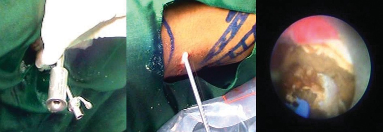
图 2 微通道经皮肾镜鞘管就位(左)、正在进行肾镜检查(中)以及采用钬激光的体内碎石(右)
PCNL 的缺点
PCNL 的并发症发生率为15%~39%。最常见的并发症是发热和失血需要输血。由于患儿的器官相对拥挤且彼此毗邻,容易出现血胸、胸腔积液或结肠损伤等并发症。然而,采用超声引导穿刺并配合细致的操作技术,可以避免这些并发症。
输尿管软镜术 (fURS/RIRS)
由于柔性输尿管镜的微型化持续进步,RIRS 现在已可在儿科患者中以极少并发症的方式实施。以往由于输尿管缺血、输尿管穿孔和狭窄形成等并发症,儿童一般避免进行 RIRS。然而,目前柔性内镜的规格已小至 7.5 Fr,能够较容易地置入儿童输尿管内。同时,直径小至 200 微米且具备良好碎石与粉末化能力的小型激光光纤的发展,使结石清除率显著提高,可达 85%至90%。并发症最多见于体重不足 20 kg 的儿童。28 AUA 指南也建议,对于小于 20 mm 的结石,推荐 URS 或 ESWL。儿童需在全身麻醉下进行 fURS。体位与成人相同,将患儿置于改良截石位,并对受压部位充分垫衬。首先,在 C 臂引导下,使用膀胱镜将导丝置入输尿管。可使用逐级扩张器将输尿管扩张至最多 10 Fr。可使用输尿管通道鞘(10/12、11/13、12/14 Fr)。在迂曲的输尿管内,通道鞘有助于推进柔性输尿管镜。若在任何部位输尿管无法容纳。输尿管不应过度扩张。常可在不使用鞘的情况下进行手术。对于非常年幼的儿童,最好进行 presenting。应注意避免损伤输尿管及尿道。
尺寸可达7.5 Fr 的一次性柔性输尿镜的出现,重新引发了人们对柔性内镜的兴趣。此外,新的激光设备不断涌现,如高功率钬激光、铥光纤激光,其宣称在粉末化与碎石方面优于传统的低功率钬激光。
fURS 的缺点
fURS 的并发症包括感染、脓毒症、出血、输尿管损伤和狭窄。如果发现输尿管损伤,则应终止手术并置入输尿管支架。
腹腔镜与开放手术的作用
AUA 和内镜泌尿学会不建议在儿科肾结石患者中常规使用开放手术、腹腔镜手术或机器人辅助手术,除非存在先天性肾盂输尿管连接部(PUJ)梗阻。28
结论
在当今时代,泌尿内镜治疗仍是小儿结石疾病的主要治疗手段。小儿肾结石虽不常见,但会给泌尿外科医生带来独特的挑战。不过,指导治疗的原则基本相同,仅需少量调整。首先且最重要的是,目标仍是以尽可能少的再次治疗次数实现结石的完全清除,从而将并发症降至最低。
由于与内镜泌尿外科相比成功率较低,以及器械的小型化与光学系统的改进,ESWL的使用已显著减少。ESWL可作为内镜治疗的辅助手段。
mini PCNL、micro PCNL 和 mini-micro PCNL 的出现,配合更为纤细的器械以及超声引导下穿刺,为在小儿患者中实现更好的接受度和更低的并发症发生率铺平了道路。
采用柔性输尿管镜的RIRS也已成为常规操作,并且由于柔性输尿管镜直径减小、成像质量提高以及用于碎石的各类激光的可获得性更高,随着时间的推移有望成为肾结石的标准治疗。
本书相关章节讨论了输尿管结石和膀胱结石的处理。
要点
- 内镜泌尿学治疗在当代仍是儿科结石疾病的主要治疗手段。儿科肾结石对泌尿外科医生提出了独特挑战。首要目标是在尽可能少的再次治疗和尽量少的并发症前提下实现完全清除结石。
- ESWL 仍是肾结石的一种治疗选择,其无石率在 68% 至 84% 之间。尽管由于其成功率低于内镜治疗,ESWL 的应用已显著减少,但它可作为内镜治疗的辅助手段。
- mini PCNL、micro PCNL 和 mini-micro PCNL 的出现,配合纤细精巧的器械与超声引导穿刺,使其在儿科患者中的接受度更高且并发症发生率更低。此外,大功率钬激光、铥激光光纤(TLF)、Trilogy 和 Shockpulse 等能量源提高了结石清除率。使用带吸引功能的鞘管具有额外优势。
- 软性输尿管肾镜如今更为纤细,可在儿科输尿管内使用。此外,与旧款相比,其光学性能也更佳。应注意避免对输尿管过度扩张。许多情况下可进行无鞘操作。
参考文献
- CD S Jr, AC S, JM H. Prevalence of kidney stones in the. . DOI: 10.1016/j.eururo.2012.03.052.
- States U. Urologic Diseases in America Project. Eur Urol 2012; 62: 160. DOI: 10.1097/01.ju.0000154031.18562.0c.
- Sas DJ, Hulsey TC, Shatat IF. Increasing incidence of kidney stones in children evaluated in the emergency department. J Pediatr 2010; 157 (1): 132–137. DOI: 10.1016/j.jpeds.2010.02.004.
- Bush NC, Xu L, Brown BJ. Hospitalizations for pediatric stone disease in United States. J Urol 2002; 183 (3): 1151–1156. DOI: 10.1016/j.juro.2009.11.057.
- Matlaga BR, Schaeffer AJ, Novak TE. Epidemiologic insights into paediatric kidney stone disease. Urol Res 2010; 38 (6): 453–457. DOI: 10.1007/s00240-010-0327-9.
- Wang HH, Wiener JS, Lipkin ME. Estimating the nationwide, hospital based economic impact of pediatric urolithiasis. J Urol 1859; 193 (5 Suppl). DOI: 10.1016/j.juro.2014.09.116.
- Lao M, Kogan BA, White MD. High recurrence rate at 5-year followup in children after upper urinary tract stone surgery. J Urol 2014; 191 (2): 440–444. DOI: 10.1016/j.juro.2013.09.021.
- Tasian GE, Kabarriti AE, Kalmus A. Kidney stone recurrence among children and adolescents. J Urol 2017; 197 (1): 246–252. DOI: 10.1016/j.juro.2016.07.090.
- Tekin A, Tekgul S, Atsu N. Oral potassium citrate treatment for idiopathic hypocitruria in children with calcium urolithiasis. J Urol 2002; 168 (6): 2572–2574. DOI: 10.1097/00005392-200212000-00076.
- Tasian GE, Cost NG, Granberg CF. Tamsulosin and spontaneous passage of ureteral stones in children: a multi-institutional cohort study. J Urol 2014; 192 (2): 506–511. DOI: 10.1016/j.juro.2014.01.091.
- Choi H, Snyder HM D III, J.W.. Urolithiasis in childhood: current management. J Pediatr Surg 1987; 22: 158–164. DOI: 10.1016/s0022-5347(17)43213-7.
- Chaussy C, Brendel W, Schmiedt E. Extracorporeally induced destruction of kidney stones by shock waves. Lancet 1980; 2: 1265–1268. DOI: 10.1016/s0140-6736(80)92335-1.
- Newman DM, Coury T, Lingeman JE. Extracorporeal shock wave lithotripsy in children. J Urol 1986; 136: 238–240. DOI: 10.5144/0256-4947.2001.97.
- AC H, JB H, EJ K. First and second generation lithotripsy in children: Results comparison and follow-up. J 1995; 153: 1969–1971. DOI: 10.1016/s0022-5347(01)67380-4.
- Lifshitz DA, Lingeman JE, Zafar FS. Alterations in predicted growth rates of pediatric kidneys treated with extracorporeal shockwave lithotripsy. J Endourol 1998; 12: 469–475. DOI: 10.1089/end.1998.12.469.
- Traxer O, Lottmann H, Archambaud F. Extracorporeal lithotripsy in children. Study of its efficacy and evaluation of renal parenchymal damage by DMSA-Tc 99m scintigraphy: a series of 39 children. Arch Pediatr 1999; 6: 251–258.
- Villanyi KK, Szekely JG, Farkas LM. Short-term changes in renal function after extracorporeal shock wave lithotripsy in children. J Urol 2001; 166: 222–224. DOI: 10.1097/00005392-200107000-00068.
- Assimos D, Krambeck A, Miller NL. Surgical management of stones: American Urological Association/Endourological Society Guideline. J Urol 2016; 196 (4): 1161–1169. DOI: 10.1016/j.juro.2016.05.090.
- Woodside S JR, GF S, G.L.. Percutaneous stone removal in children. J Urol 1985; 134 (1166). DOI: 10.1016/s0022-3468(86)80487-0.
- Raza A, Smith G, Moussa S, Tolley D. Ureteroscopy in the management of pediatric urinary tract calculi. J Endourol 2005; 19: 151–158. DOI: 10.1089/end.2005.19.151.
- Desai MR, Kukreja RA, Patel SN, Bapat SD. Percutaneous nephrolithotomy for complex pediatric renal calculus disease. J Endourol 2004; 18: 23–27. DOI: 10.1089/089277904322836613.
- Samad L, Aquil S, Zaidi Z. Paediatric percutaneous nephrolithotomy: setting new frontiers. BJU Int 2006; 97: 359–363. DOI: 10.1111/j.1464-410x.2006.05932.x.
- Berrettini A, Boeri L, Montanari E. Retrograde intrarenal surgery using ureteral access sheaths is a safe and effective treatment for renal stones in children weighing <20 kg. J Pediatr Urol 2018; 14 (1). DOI: 10.1016/j.jpurol.2017.09.011.
- Mahmud M, Zaidi Z. Percutaneous nephrolithotomy in children before school age: experience of a Pakistani centre. BJU Int 2004; 94: 1352–1354. DOI: 10.1111/j.1464-410x.2004.05173.x.
- Zeren S, Satar N, Bayazit Y, Bayazit AK, Payasli K, Ozkeçeli R. Percutaneous nephrolithotomy in the management of pediatric renal calculi. J Endourol 2002; 16: 75–78. DOI: 10.1089/089277902753619546.
- Manohar T, Ganpule AP, Shrivastav P, Desai M. Percutaneous nephrolithotomy for complex calyceal calculi and staghorn stones in children less than 5years of age. J Endourol 2006; 20: 547–551. DOI: 10.1089/end.2006.20.547.
- Salah MA, Tóth C, Khan AM, Holman E. Percutaneous nephrolithotomy in children: Experience with 138 cases in a developing country. World J Urol 2004; 22: 277–280. DOI: 10.1007/s00345-004-0454-4.
- Nouralizadeh A, Basiri A, Javaherforooshzadeh A, Soltani MH, Tajali F. Experience of percutaneous nephrolithotomy using adult sized instruments in children less than 5 years old. J Pediatr Urol 2009; 5: 351–354. DOI: 10.1016/j.jpurol.2008.12.009.
- Ganpule AP, Mishra S, Desai MR. Percutaneous nephrolithotomy for pediatric urolithiasis. Indian Journal of Urology: IJU: Journal of The Urological Society of India 2010; Oct;26(4):549. DOI: 10.4103/0970-1591.74458.
最近更新时间: 2025-09-22 08:00